Vitamin D, also known as the sun vitamin, is a hormone which has long been known to regulate calcium and phosphorus homeostasis, and protect the integrity of the skeletal system [1]. It has been shown that the skin is not only the site where vitamin D biosynthesis is triggered, but also the target organ for the biologically active form of this vitamin. It is implicated in a wide array of skin functions, ranging from the proliferation, differentiation and apoptosis of keratinocytes to the maintenance of the protective barrier and immunoregulatory processes. Vitamin D administration is a therapeutic option in the management of a number of skin conditions. A link has been found between vitamin D levels and the pathogenesis of psoriasis, atopic dermatitis (AD), but also melanoma or excessive hair loss [2].
Vitamin D synthesis in the skin
The main source of vitamin D for the body is the epidermis. In response to sunlight (UV radiation with wavelengths in the range of 280-320 nm; UVB), 7-dehydrocholesterol in the epidermis is converted into vitamin D which – in the presence of enzymes (25-hydroxylase and 1-hydroxylase) present in epidermal cells (keranocytes) – is metabolised to the active metabolite (calcitriol; 1,25(OH)2D3). Like most other cells, keratinocytes also have vitamin D receptors (VDR), so they are able to respond to the generated 1,25(OH)2D3 [3].
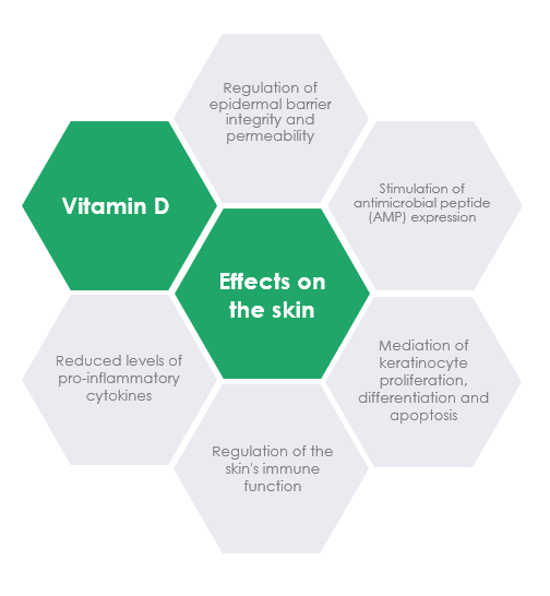
Role of vitamin D in the skin
Epidermal differentiation and proliferation
Vitamin D, through its effects on epithelial cell proliferation and differentiation, modulation of the immune system and the process of apoptosis, plays a major role in the homeostasis of the skin’s barrier function. Multiple in vitro and in vivo studies have demonstrated a dose-dependent effect of vitamin D on the proliferation and differentiation of keratinocytes. Interestingly, low concentrations of vitamin D promote the proliferation of keratinocytes, but at higher pharmacological doses a distinct inhibitory effect is observed [4]. Studies have shown that 1,25(OH)2D3 regulates the proliferation of keratinocytes in the epidermal basal layer, enhances the synthesis of keratin, and regulates the synthesis of filaggrin, loricrin, involucrin and transglutaminase – substances implicated in the differentiation of keratinocytes into cells of the stratum corneum with a dense structure, resistant to chemical and physical factors, serving as the “backbone” of the epidermal barrier. In addition, vitamin D helps regulate the synthesis of glucosylceramides necessary to maintain the integrity of the stratum corneum, which further strengthens the epidermal barrier. A decreased level or deficiency of 1,25(OH)2D3 is known to impair epidermal differentiation, resulting in excessive proliferation of the basal layer and contributing to the pathomechanism of psoriasis [2]. Studies have found that the topical application of calcitriol restores the epidermal barrier and produces an antibacterial effect on the skin damaged by treatment with corticosteroids or sodium lauryl sulphate [5].
Vitamin D is also involved in the apoptosis of keratinocytes. At physiological levels, it prevents apoptosis caused by various proapoptotic factors (e.g. UV radiation), while at high concentrations, it induces apoptosis in keratinocytes. The latter process is used in the treatment of psoriasis, among other conditions (Fig. 1) [6].
Effect on the skin’s immune activity
The skin’s innate immunity consists of physical barrier structures such as the stratum corneum, immune cells (neutrophils, monocytes, macrophages, dendritic cells, NK cells, etc.) and antimicrobial peptides (AMPs). The process of cutaneous synthesis of AMPs is the skin’s primary protective mechanism against microbial invasion. Skin cells – including keratinocytes, sebaceous cells, eccrine gland cells and mast cells – contribute to the synthesis of AMPs, primarily β-defensin and cathelicidin, in the skin [7]. In addition to their antibacterial, antifungal and antiviral properties, these peptides also regulate the immune system, participating in processes including cytokine and chemokine release, antigen presentation, cell proliferation, vascular permeability, angiogenesis, and wound healing. AMP levels are low in intact skin, but skin injury and infection result in an increased synthesis of 1α-hydroxylase, which causes a rise in the local production of active vitamin D and leads to a local increase in AMP generation.
In addition to mediating the synthesis of AMPs in the skin, 1,25(OH)2D3 regulates the skin’s immune response by reducing antigen presentation and modulating the process of cytokine production by keratinocytes (Fig. 1) [8].
Role of vitamin D in some inflammatory skin diseases
Psoriasis
Maintaining adequate vitamin D levels plays a key role in relieving skin lesions and treating psoriasis. In addition to modulating or suppressing inflammation, vitamin D administered in high doses is known to inhibit excessive proliferation of the epidermis and induce keratinocyte apoptosis, which in turn reduces psoriatic plaque growth.
Case-control studies have revealed significantly lower serum levels of 25(OH)D3 in patients with psoriasis compared to the control group, and an inverse correlation between 25(OH)D3 levels and disease severity [6]. Also, extensive research shows that vitamin D may be effective in relieving the symptoms of psoriasis. Psoriasis patients who received 35,000 IU of vitamin D3 once daily for 6 months showed a significant improvement in their PASI (psoriasis area severity index) scores, and a clear increase in the serum levels of 25(OH)D3 [9].
The clinical improvement noted after phototherapy in patients with psoriasis is believed to be indirectly due to an increase in 25(OH)D3 levels in the skin [6]. Also, combinations of vitamin D or its analogues with a corticosteroid have been shown to be more effective than monotherapy because of their complementary characteristics. In combination therapy, vitamin D may counteract steroid-induced skin atrophy by restoring the epidermal barrier, while a corticosteroid may relieve skin irritation induced by vitamin D analogues [10].
Atopic dermatitis (AD)
AD is a chronic recurrent skin disease arising from complex interactions between genetic, immunological and environmental factors; it is characterised by chronic inflammation, damage to the epidermal barrier, immune disturbances, and elevated serum IgE levels.
Large population studies have provided evidence for an increased risk of AD development in individuals with vitamin D deficiency or inadequate vitamin D status. Serum vitamin D levels have been shown to be lower in AD patients than in the controls, which points towards an association between vitamin D deficiency and the risk of atopic eczema. The severity of AD has also been found to be inversely correlated with vitamin D levels. Lower levels of vitamin D were detected in patient groups with moderate to severe AD compared to patients with mild AD [6]. Also, low maternal vitamin D levels during pregnancy increase the risk of disease in infants early in life [11].
A metaanalysis of clinical trials has shown that vitamin D supplementation significantly reduces the severity of AD symptoms [12, 13], which is probably attributable to the restoration of normal immune function [13].
AD is also associated with an elevated IgE response to common environmental and food allergens. Vitamin D is known to have an inhibitory effect on the allergic response: vitamin D administration has been found to inhibit the production of IgE by human B cells and suppress IgE-mediated activation of mast cells both in vitro and in vivo [14].
Patients with AD are prone to skin colonisation and infection with a variety of pathogens including Staphylococcus aureus and the Herpes simplex virus. A clinical trial showed that oral vitamin D supplementation (2,000 IU/day) for 4 weeks reduced skin colonisation by S. aureus and relieved clinical symptoms in AD patients [15]. In another study, clinical improvement was achieved by administering vitamin D to children with atopic dermatitis and eczema herpeticum at a dose of 6,000-10,000 IU/day (depending on patient age and deficiency status) for 2 months [16].
Ensuring an adequate vitamin D status in the body in patients with AD restores balance in the immune system dysregulated by the disease, repairs defects in epidermal barrier, and increases the production of antibacterial peptides which prevent pathogenic colonisation.
Conclusions
Since vitamin D has multifaceted effects on the skin, maintaining an appropriate vitamin D status is essential for proper skin function. By strengthening the epidermal barrier, vitamin D regulates cell divisions and apoptosis of skin cells, and balances immune activity. In addition, it is an element of a major therapeutic strategy for the management of multiple skin conditions, mainly psoriasis and atopic dermatitis.
REFERENCES
Holick MF. N Engl J Med. 2007;357:266–281
Barrea L, Savanelli MC, Di Somma C, et al. Rev Endocr Metab Disord. 2017;18(2):195-205.
Kim MJ, Kim SN, Lee YW, et al. Nutrients 2016;8:E789.
Di Filippo P, Scaparrotta A, Rapino D, et al. Int Arch Allergy Immunol 2015;166:91-96.
Yip KH, Kolesnikoff N, Yu C, et al. J Allergy Clin Immunol 2014;133:1356-1364, e1351-e1314.
Udompataikul M, Huajai S, Chalermchai T, et al. J Med Assoc Thai 2015;98(suppl 9):S23-S30.
Albenali LH, Danby S, Moustafa M, et al. J Allergy Clin Immunol 2016;138:1715-1719. e1714.
BIOGRAPHICAL NOTE
Dr n. farm. Marlena Dudek-Makuch,
Ekspert ds. Rozwoju w Curtis Health Caps, Wysogotowo.
Marlena Dudek-Makuch, PhD (Pharm.), MSc, has 20 years of professional experience in phytochemical and biological research, and scientific information (assistant professor at the Department of Pharmacognosy, Poznań University of Medical Sciences). She is an author of research and review papers on the isolation and identification of compounds of plant origin, and evaluation of their biological activity. Since 2015, she has been teaching a postgraduate course in “Herbs in practice and therapy”.
She is currently a member of the Regulatory Department, R&D Division, at CHC. In her position, she is in charge of preparing expert reports (clinical, non-clinical) for medicinal products, clinical reports for OTC switches, clinical assessments of medical devices, pharmacovigilance activities for medical devices, and safety assessment of plant-based raw materials used in medicinal products, medical devices, and dietary supplements.